
Product Center
Your Location : Home / Product Center / Ophthalmic Equipments / Ophthalmic Ultrasound AB Scan+ Pachymeter / SW-2100 Ophthalmic AB Scan
| Name: | SW-2100 Ophthalmic AB Scan |
|---|---|
Product Description
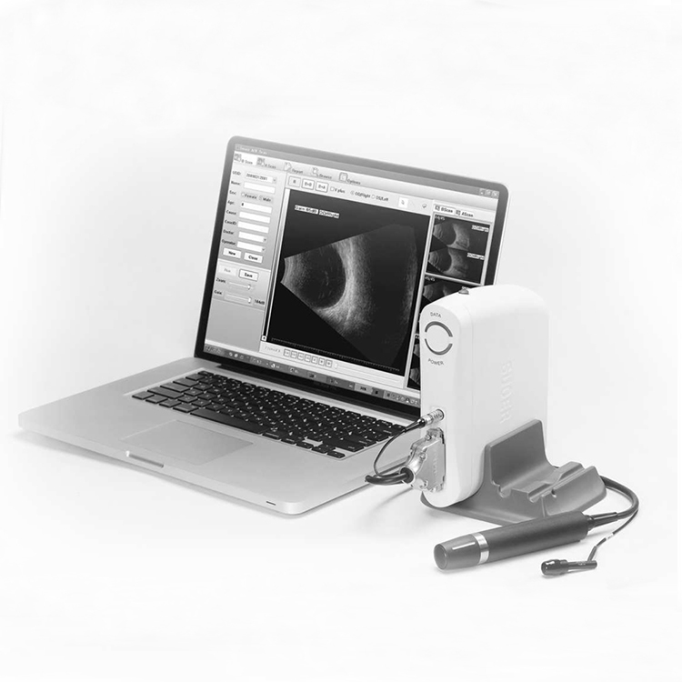
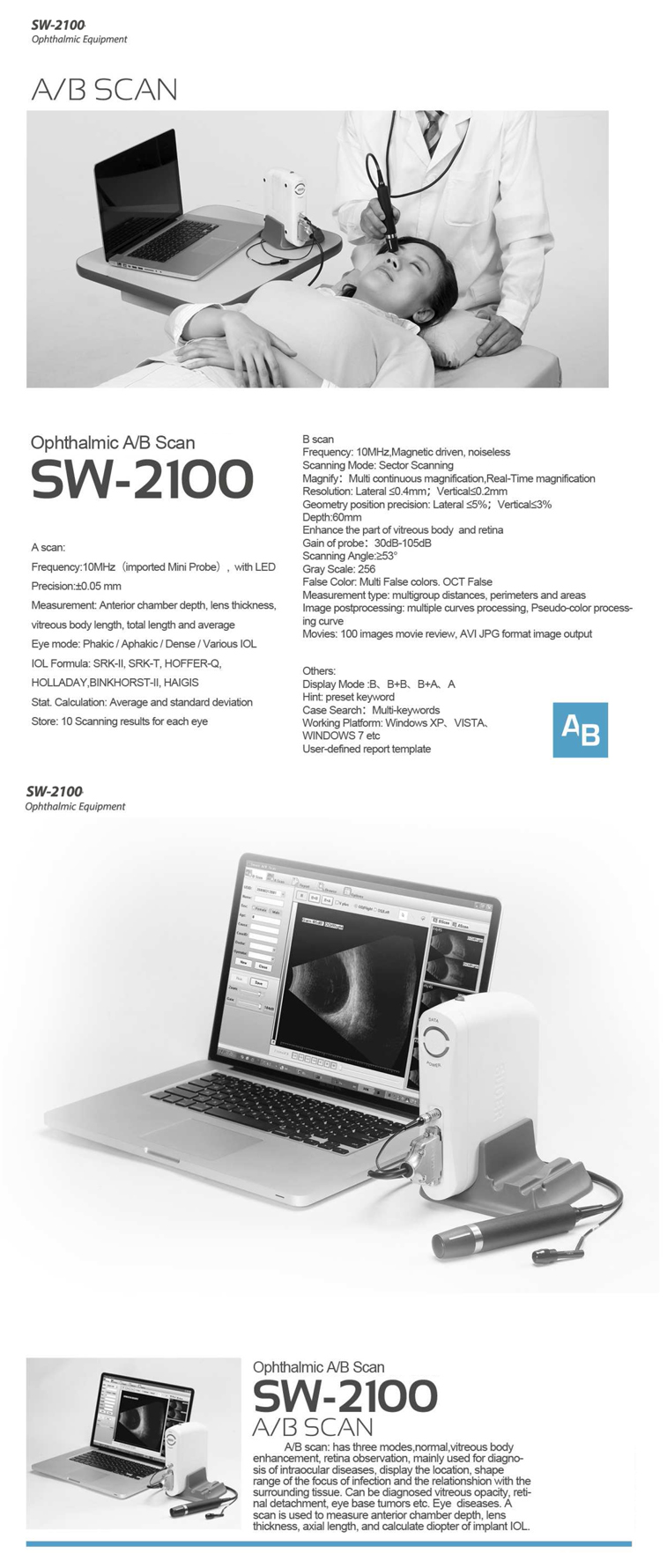
SW-2100 Ophthalmic AB Scan
Features:
AB scan: has three modes, normal, vitreous body enhancement, retina observation, mainly used for diagnosis of intraocular
diseases, display the location, shape range of the focus of infection and the relationship with the surrounding tissue.
Can be diagnosed vitreous opacity, retinal detachment, eye base tumors etc.
Eye diseases. A scan is used to measure anterior chamber depth, lens thickness, axial length, and calculate diopter of implant IOL.
Parameters:
A scan | |
Frequency: | 10MHz(imported Mini Probe, with LED |
Precision: | ±0.05 mm |
Measurement | Anterior chamber depth, lens thickness, |
Measurement: | AL, ACD, L, V and the average vitreous body length, total length and average |
IOL Formula: | SRK-II, SRK-T, HOFFER-Q, HOLLADAY,BINKHORST-II, HAIGIS |
Stat Calculation: | Average and standard deviation |
Store: | 10 Scanning results for each eye |
B scan | |
Frequency: | 10MHz ,Magnetic driven, noiseless |
Scanning Mode: | Sector Scanning |
Magnify: | Multi continuous magnification,Real-Time magnification |
Resolution: | Lateral ≤0.4mm;Vertical≤0.2mm |
Depth: | 60mm |
Gain of probe | 30dB-105dB |
Scanning Angle: | 53° |
Gray Scale: | 256 |
False Color: | Multi False colors. OTC False |
Measurement type | Multi-group distances, perimeters and areas |
Image post processing | multiple curves processing, Pseudo-color processing curve |
Movies: | 100 images movie review,AVI ZIP JPG format image output |
Other | |
Display Mode: | B,B+B,B+A,A |
Hint | preset keyword |
Case Search: | Multi-keywords |
Working Platform | Windows XP、VISTA、WINDOWS 7 etc |
User-defined report | template |


 AB Scan")