
Product Center
Your Location : Home / Product Center / Ophthalmic Equipments / Optical Coherence Tomography / OSE-4000 Optical Coherence Tomography
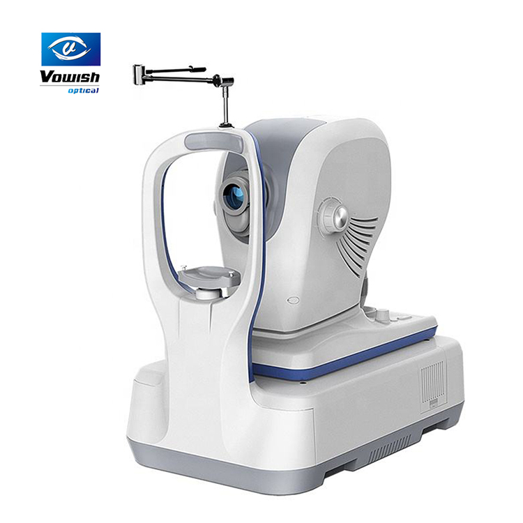
| Name: | OSE-4000 Optical Coherence Tomography |
|---|---|
Product Description
OSE-4000 Optical Coherence Tomography
Features:
Macula Six-line Radial:Have a glimpse of the retina via HD imaging and quick data analysis
Macula Multi: Multiple HD cross-sectional images acquisition
Macula Cube: A point-by point assessment of retinal thickness with a 500 x 100 dense cube
Anterior HD line: High definition OCT imaging of the cornea enables localization of the Bowman's layer, the interface between corneal stroma and epithelium
Anterior Six-line Radial: The anterior segment scanning through 6 radial lines of equal length can be used to measure the central corneal thickness
VASC ANM OCT ANGIOGRAPHY NEW: Valuable OCTA for routine clinical practice Optical Coherence Tomography Angiography (OCTA) is a new
non-invasive imaging technique that allows the detailed study of flow within the vascular structure of the eye without the need of dye injections.
HD SLO + EYE TRACKING
Parameters:
OCT Imaging | Methodology | Spectral domain OCT | |
Optical source | Super luminescent diode (SLD), 840 nm | ||
Scan speed | 80,000 A-scans/s | ||
Axial resolution (optical) | 5 microns (optical), 3.6 microns (digital) | ||
Transverse resolution | 15 microns (optical), 3 microns (digital) | ||
A-scan depth | 3mm | ||
Diopter range | - 20 to + 20 diopters | ||
Scan patterns | Macular: HD line scan (6 / 12 mm), 3D scan (6 mm x 6 mm), 6-line radial scan, Multi (X-Y: 5x 5) Disc:3Dscan(6mmx6mm) Anterior: HD line scan (6 / 16mm), 6-line radial scan | ||
Fundus Imaging | Methodology | Line scanning laser ophthalmoscopy (LSLO) | |
Minimum pupil diameter | 3.0 mm | ||
Field of view | 45 degrees | ||
VASCANTM OCTA MODULE | Scanning volume/area | VASCAN Advance | VASCAN Essential |
3mmx3mm 256x256 A-scans 6mmx6mm 360x360 A-scans 8mmx8mm 360x360 A-scans 12mmx8mm 540x360 A-scans | 3mmx3mm 256x256 A-scans 12mmx8mm 540x360 A-scans | ||
Algorithm | C-OMAG | C-OMAG | |
Segmentation options | Encoded, Vitreousretina Intrerface(VRI), Superfcial retina, Deepfcial retinal, Avascular, Choriocapillaris, Choriod, Custom | ||
Quantitative analysis | Yes | Not available | |
EL ECTRICAL AND PHYSICAL | Weight | 30.5kg | |
Dimension | 532mm(L)x360mm(W)x540mm(H) | ||
Source voltage | AC 100-240V, 50Hz-60Hz | ||
Power input | 90VA | ||


